Return
- Core Technology
- Based on the foundation of proprietary technologies and SenseCore AI infrastructure,
SenseTime has rapidly opened up AI application in multiple vertical scenarios, and is empowering various industries.
- 01Multi-modality Image Registration
- 02Lesion/Body Part Segmentation and Quantitative Analysis
- 03Differentiation of Benign and Malignancy & Grading of Disease
- 04Lesion Classification
- 05Focus/nidus detection and locating
01 / 05
Multi-modality Image Registration
With the registration of multi-modality data such as CT, MRI, and PET of the same body part or organ, the system can achieve accurate fusion of different modalities and different sequence data, enabling more accurate precise qualitative grading and quantitative analysis of lesions.

02 / 05
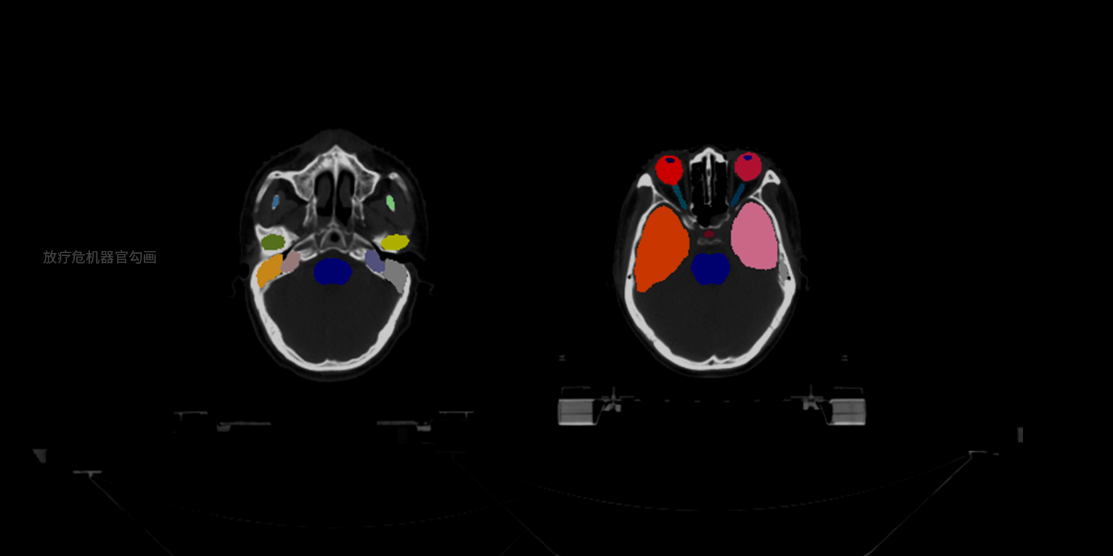
Lesion/Body Part Segmentation and Quantitative Analysis
The system supports small dataset training on medical images to conduct pixel-level precise segmentation of multiple lesions and organs, and automatically carry out quantitative analysis of key parameter measurement, such as radiotherapy target area delineation and pelvic tumor segmentation. The system does not only release doctors from time-consuming, labor-intensive manual illustration work, but also assist doctors in quantitative diagnosis and personalized surgical planning.
03 / 05
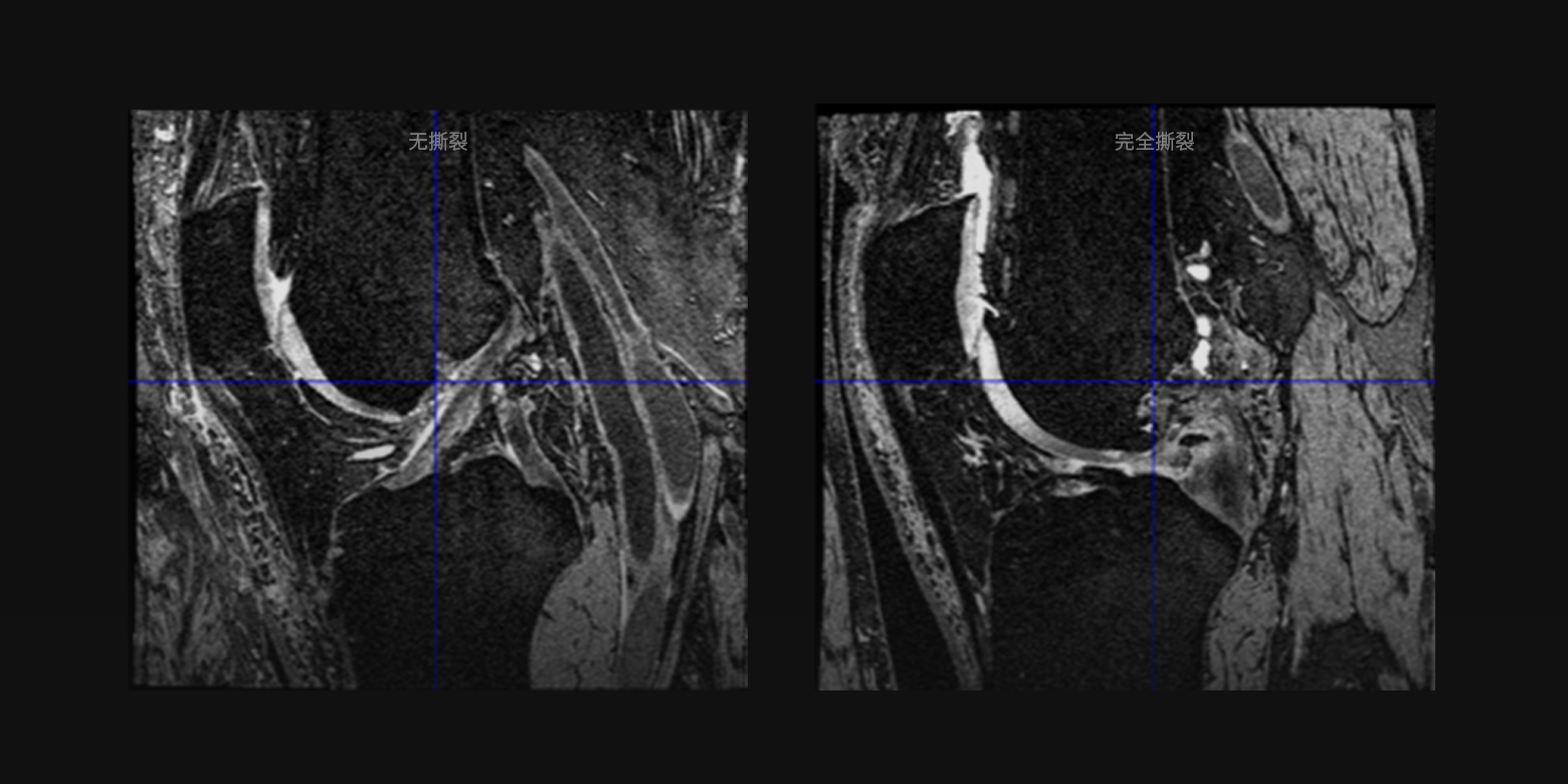
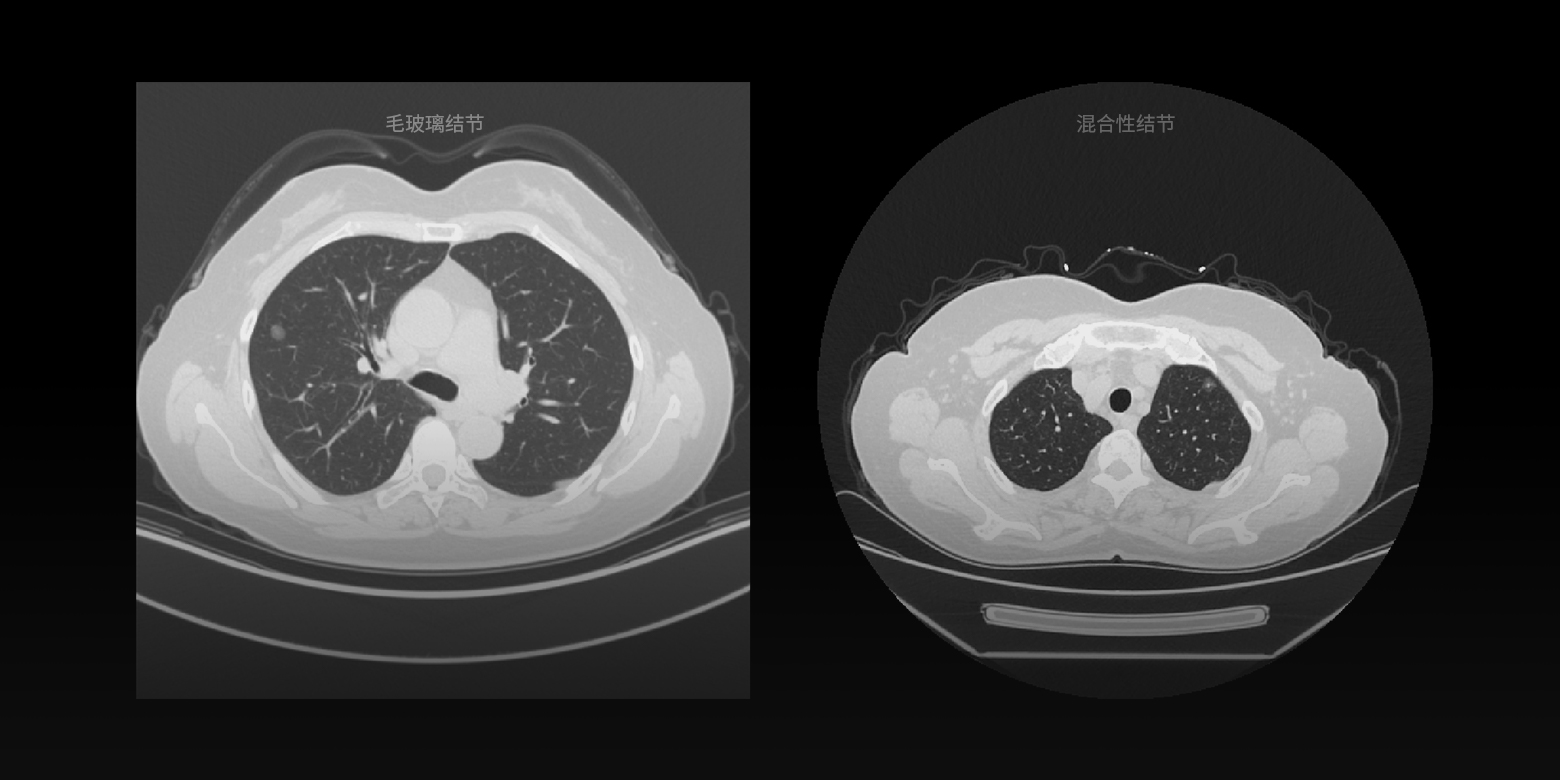
Differentiation of Benign and Malignancy & Grading of Disease
Based on the analysis of medical images and clinical factors, the system can diagnose the malignancy and severity of various diseases, such as grading the severity of anterior cruciate ligament tear, differentiating between benign and malignant lung nodules, in order to optimize the process of mass screening and hierarchical diagnosis.
04 / 05
Lesion Classification
Based on international diagnostic guidelines and the experience of top medical professionals, the system swiftly and precisely classifies lesions, which plays a key role in clinical diagnosis and helps minimize misdiagnosis of similar lesions.
05 / 05
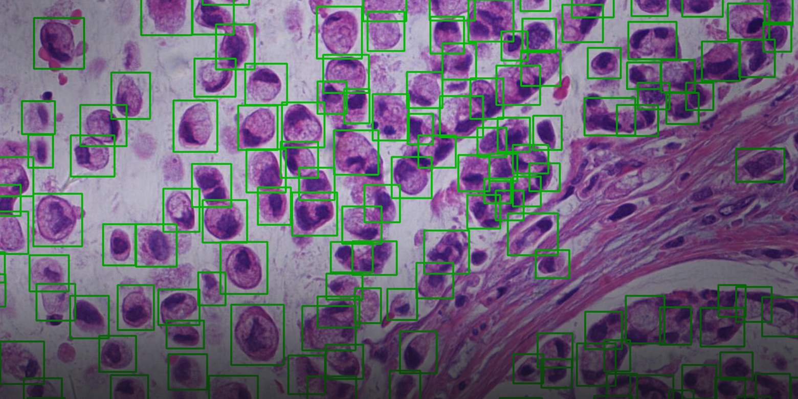
Focus/nidus detection and locating
Employing cutting-edge computer vision technology, we can fast detect and locate various focuses and nidi (for example, cell detection and locating based on pathological images) found through multimodality imaging such as computed tomography (CT), magnetic resonance imaging (MRI) and digital pathology. These services allow physicians to make informed decisions in diagnostic tests, and make the diagnosis process more efficient.
01 / 05
Multi-modality Image Registration
With the registration of multi-modality data such as CT, MRI, and PET of the same body part or organ, the system can achieve accurate fusion of different modalities and different sequence data, enabling more accurate precise qualitative grading and quantitative analysis of lesions.
02 / 05
Lesion/Body Part Segmentation and Quantitative Analysis
The system supports small dataset training on medical images to conduct pixel-level precise segmentation of multiple lesions and organs, and automatically carry out quantitative analysis of key parameter measurement, such as radiotherapy target area delineation and pelvic tumor segmentation. The system does not only release doctors from time-consuming, labor-intensive manual illustration work, but also assist doctors in quantitative diagnosis and personalized surgical planning.
03 / 05
Differentiation of Benign and Malignancy & Grading of Disease
Based on the analysis of medical images and clinical factors, the system can diagnose the malignancy and severity of various diseases, such as grading the severity of anterior cruciate ligament tear, differentiating between benign and malignant lung nodules, in order to optimize the process of mass screening and hierarchical diagnosis.
04 / 05
Lesion Classification
Based on international diagnostic guidelines and the experience of top medical professionals, the system swiftly and precisely classifies lesions, which plays a key role in clinical diagnosis and helps minimize misdiagnosis of similar lesions.
05 / 05
Focus/nidus detection and locating
Employing cutting-edge computer vision technology, we can fast detect and locate various focuses and nidi (for example, cell detection and locating based on pathological images) found through multimodality imaging such as computed tomography (CT), magnetic resonance imaging (MRI) and digital pathology. These services allow physicians to make informed decisions in diagnostic tests, and make the diagnosis process more efficient.